Most people think an eye exam is just about checking if you need glasses. But a complete eye exam does much more than that. It looks at the health of the structures inside your eye, including the retina.
The retina is the light-sensitive tissue at the back of your eye. It is where early signs of serious eye disease often appear first. Seeing it clearly matters.
Optomap retinal imaging gives your eye doctor a detailed, wide view of your retina in seconds. No drops. No blurry vision. No waiting around. It is one of the most useful tools in modern preventive eye care.
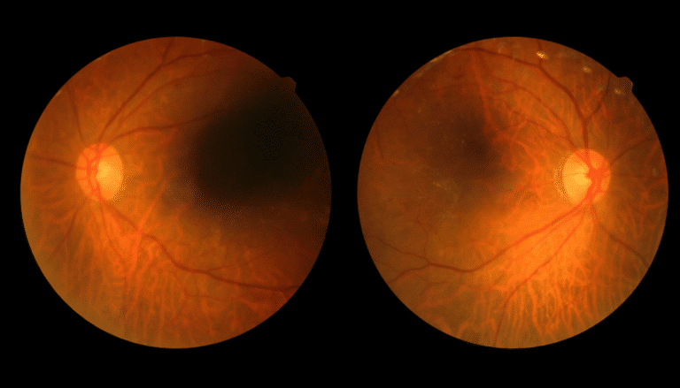
Optomap is an ultra-widefield retinal imaging technology developed by Optos. It captures up to 200 degrees of your retina in a single scan. That is roughly 80% of the entire retina.
A standard eye exam without any imaging only lets the doctor view a small portion of the retina at a time. Optomap changes that. It gives a much broader picture in far less time.
The scan is quick, painless, and non-invasive. You simply look into the device for a brief moment, and the image is captured in under one second.
Book your Vision Rehabilitation today and experience care that puts your eyes first.
One of the biggest reasons patients appreciate Optomap is that it does not require dilating eye drops. Traditional dilation causes blurry vision for several hours and makes your eyes very sensitive to light. With Optomap, you walk out of the office seeing normally, ready to drive and get on with your day.
Standard retinal viewing through an ophthalmoscope shows roughly 10 to 12% of the retina at a time. Optomap captures up to 80% in one image. This wider view means more of the retina is screened in a single scan, including the peripheral areas where some conditions first appear.
The scan itself takes less than a second. The full process, including positioning and image review, adds only a few minutes to your appointment. There are no bright lights, no prolonged exposure, and no discomfort.
Your Optomap image is stored digitally in your file. At your next visit, your doctor can compare the new image with your previous one. This makes it easier to spot subtle changes over time. Tracking those changes is one of the best ways to catch disease early.
Optomap helps identify a range of eye conditions and systemic health issues that show up in the retina.
The retina is one of the few places in the body where blood vessels can be seen directly and without surgery. That makes it a valuable window into your overall health.
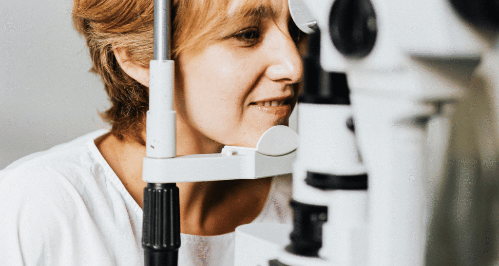
Positioning You sit comfortably in front of the Optomap device. You rest your chin on a support and bring your eye close to the opening. The process is simple and takes no preparation.
Image Capture You look at a small target light inside the device. The scan captures your retinal image in under one second. Most patients experience nothing more than a brief, gentle flash of light.
Review Your eye doctor reviews the image immediately and discusses the findings with you. If previous scans are on file, they are compared side by side. You can often see your own retina on a screen during this conversation.
Many patients wonder how Optomap compares to getting their eyes dilated. Here is a straightforward comparison:
Feature | Optomap Imaging | Traditional Dilation |
Time needed | Seconds | 20 to 40 minutes |
Comfort level | Painless, no drops | Blurry vision and light sensitivity |
Field of view | Up to 200 degrees | Around 45 degrees |
After-effects | None | Recovery time needed |
View type | Wide peripheral retina | Detailed central retina |
The key point here is that these two methods are not competing with each other. They are complementary. Optomap gives a wide view across the whole retina. Dilation allows your doctor to look more closely at specific areas in the center.
Many eye doctors use both during the same exam for the most complete evaluation possible.
Optomap is typically an out-of-pocket add-on to your standard eye exam. Most vision insurance plans do not cover it because it is considered an elective screening upgrade.
Pricing varies by clinic. The cost is generally modest given the level of information it provides. Many patients find the convenience alone worth it, particularly those who want to avoid the side effects of dilation.
If you are unsure whether your plan covers any portion of it, check with your insurance provider. Some medical insurance plans may cover Optomap when it is used to monitor a diagnosed condition like diabetic retinopathy or glaucoma.
Optomap is an excellent screening tool, but it does have limitations worth understanding.
Consider adding Optomap to your exam if:
For patients managing conditions like eye conditions that affect vision long-term, annual Optomap imaging is a practical way to track changes before symptoms appear.
Optomap is a quick, non-invasive scan that captures up to 200 degrees of the retina in less than a second. It is used during eye exams to screen for retinal disease and monitor eye health over time.
They serve different purposes. Optomap captures a wider view quickly and without drops. Dilation provides more detailed central retinal examination. Many doctors use both for a complete evaluation.
It is typically an out-of-pocket cost added to your regular exam fee. Pricing varies by clinic. Insurance often does not cover it unless it is medically indicated.
Most vision plans do not cover it as a routine add-on. Some medical plans may cover it if used to monitor a specific diagnosis like diabetic retinopathy or glaucoma.
Yes. It is completely safe, non-invasive, and does not use radiation. It is suitable for adults and children alike.
It can help detect glaucoma, macular degeneration, retinal tears, detachment, diabetic retinopathy, and vascular changes related to conditions like high blood pressure.
The scan itself takes under one second. Including positioning and image review, it adds only a few minutes to your appointment.
Sometimes. If your doctor sees something that needs a closer look, dilation may be recommended. In many routine exams, Optomap reduces or eliminates the need for dilation.
Looking for expert eye care, glasses, or vision therapy? Fill out the form and our team will get back to you quickly to schedule your appointment.
Give us a call to schedule your next appointment